
USG full form :
USG stands for Ultrasound Sonography in the medical field. High-frequency sound waves are used in this non-invasive imaging method to provide images of inside body components, organs, blood vessels, and other tissues.
USG – A Medical Concept Simplified
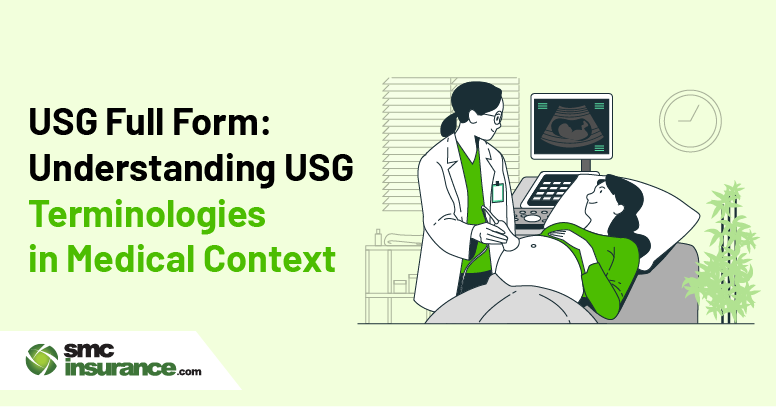
A USG, commonly known as an ultrasound, is a widely used non-invasive medical imaging technique that uses high-frequency sound waves to generate images of the inside organs and tissues of the body. In order to create real-time photographs, a transducer (probe) sends sound waves into the body and detects the returned echoes. The transducer then transmits this data to a computer and the images produced are called Sonograms. It helps with internal organ visualization, condition diagnosis, operation guidance and even therapeutic uses.
| Evolution of USG | |
|---|---|
1800s to 1940s
|
1940s to 1970s
|
1980s to 2000s
|
2010s to Present
|
Future of USG
|
|
Evolution of USG – Timeline Explained
➔ 1800s to 1940s:
- Ultrasonography’s origin rooted in physics and warfare rather than in medicine.
- Sound waves were created and detected thanks to the discovery of the piezoelectric effect in the late 1800s by researchers like Pierre Curie.
- Sonar technology was created during World Wars I and II to use sound to find submarines. These same principles were eventually applied to see inside the human body.
➔ 1940s to 1970s:
- Austrian neurologist Karl Dussik examined the brain with ultrasonography in 1942.
- Obstetric ultrasound originated in Scotland in 1958 when Ian Donald, currently regarded as the Father of Medical Ultrasound, used it to observe a pregnancy.
- Real-time imaging replaced static images.
- Physicians were able to see inside body shapes with the first B-mode (Brightness Mode) scanners. Comparing with black-and-white blobs, grayscale imaging allowed for better interpretation.
- Applications spread to the liver, heart, abdomen, and other areas.
➔ 1980s to 2000s:
- Real-time 2D imaging became commonplace.
- With the advent of Doppler ultrasonography, medical professionals could now observe blood flow and identify vascular diseases.
- Ultrasound has evolved from large, stationary devices to small, Portable devices began to show up in emergency rooms and clinics.
- 3D and 4D ultrasounds became popular, particularly for its ability to display real-time fetal movements.
- The applications of ultrasound expanded beyond radiology to include emergency medicine, anesthesiology, and even sports medicine.
➔ 2010s to Present and Future:
- Smartphones are compatible with contemporary handheld ultrasounds like the GE Vscan and Butterfly iQ.
- Artificial intelligence began to aid in the interpretation of images.
- In emergency rooms and intensive care units, point-of-care ultrasonography (POCUS) has become a crucial tool for bedside diagnostics.
- Tele-Ultrasound, Fusion Imaging and Elastography are advancing quickly with Artificial Intelligence.
Application of USG in various medical areas:
- Gastroenterology: Gastrointestinal (GI) Ultrasound is used to diagnose numerous abdominal issues like Acute Abdominal Pain, Inflammatory Bowel Disease (IBD), Gastroentritis and several functional disorders like Dysphagia, Dyspepsia and Reflux.
- Cardiology: Echocardiography (ECG) is used to diagnose a variety of cardiovascular disorders including heart failure, valve problems, heart murmurs, and coronary artery disease by evaluating the anatomy and physiology of the heart.
- Gynecology: Transvaginal Ultrasound and Abdominal Ultrasound are used in diagnosing and monitoring infertility, detecting pelvic masses and tumors, assessing ectopic pregnancy, evaluating pelvic pain, assessing ovarian dysfunction, monitoring fetal development and diagnosing fetal deformities.
- Urology: Used in assessing multiple Urinary Tract Conditions. Urosonography is used in children to diagnose Vesicoureteral Reflux (VUR).
- Therapeutic Uses: Ultrasound therapy is used in pain management, inflammation reduction, soft tissue injuries, scar tissue softening and transdermal drug delivery in rare instances.
Pros of Ultrasound:
- Painless & Non-invasive: Only a probe and little gel are required; there are no cuts, needles, or discomfort.
- Radiation-Free: Safe for all, including infants and expectant mothers.
- Real-Time Imaging: On the screen, you can see live movements of organs, blood, or even a fetus.
- Portable & Accessible: Clinics, ambulances, and isolated locations can all use this small equipment.
- Economical and Multiple usage: Far less expensive than MRI or CT scans and beneficial for the heart, muscles, blood vessels, abdomen, pregnancy, and more.
Cons of Ultrasound:
- Dependent on the Operator: The competence of the individual performing has a significant impact on the quality of the output.
- Limited Clarity & Depth: It is not ideal for imaging through bone or air (such as the brain or lungs) or for very deep structures.
- Not Always Precise: Could ignore small tumors or subtle alterations and frequently need follow-up CT/MRI.
- Variations in Image Quality: The image may be distorted by obesity or excessive amounts of gas in the abdomen.
How is USG Related to Health Insurance?
In India, most health insurance policies have diagnostic tests, including ultrasound (USG) scans, covered under them, particularly when the doctor prescribes them as part of inpatient or outpatient care. In case you're getting a USG to detect or track a medical condition—like kidney stones, gallbladder problems, or pregnancy-related complications—it might be covered under your insurance, based on your policy.
➔ Knowing USG terminologies assists policyholders:
- Understand the purpose of the test and how it is useful in treatment.
- Understand how to communicate with doctors and insurers in case of a claim.
- Maintain proper documentation at the time of filing cashless or reimbursement claims.
- Check whether their plan includes preventive health check-ups or outpatient consultations, which usually involve USG scans.
At SMCInsurance.com, we make you aware of what's covered under your health plan—such as diagnostics like USG—so that you don't worry financially while making medical choices. To check if your USG scan is covered, contact your SMC Insurance advisor

Conclusion:
From the first origin of sonar technology, ultrasonography has gone forward to become one of the most important tools in contemporary medicine. Its real -time, radiation -free imaging skills have made it inevitable in a wide range of properties, from leading emergency treatments to monitoring conception. As any tool, the effect of ultrasound varies depending on the user's skill level and conditions, but all things are considered, it has earned its reputation as a safe, reliable window in the body.
Disclaimer:
This page's content is generic and provided purely for educational and clarifying reasons. It is based on a number of online secondary sources and is subject to change. Kindly seek advice from an expert prior to making any choices.


